Home > AGS Laboratories News > Under the Scope – Cross Polarized Filters as a Clue to Diamond Origin
Under the Scope – Cross Polarized Filters as a Clue to Diamond Origin
By Wade Abel, CG, Director of Gemological Services, AGS Laboratories, and Randall Lightfoot, CGA, Gemologist Appraiser, Christopher’s Fine Jewelry
Diamond testing is one of the most important steps in the process for a diamond grading lab. The disclosures of treatment and diamond origin (natural or laboratory grown) are required; the ability to accurately report these is paramount. At AGS Laboratories, advanced detection instruments are used for these determinations. Without access to this technology, there may be some ambiguity; however, there are ways to gather clues to a diamond’s origin without advanced instrumentation.
One tool that can provide clues to a diamond’s origin, either natural or laboratory grown, is as simple as cross-polarized filters (CPF) used in a microscope. What is seen with this technique are growth patterns or strain resulting from: plastic deformation, elastic deformation surrounding inclusions, uneven distribution of defect centers and trace elements, crystal lattice dislocations, and growth striations. When CPF is used, patterns are visible that are not normally seen without CPF. Additionally, beautiful colors can be observed.
Wade Abel, CG
Randall Lightfoot, CGA
When viewed using CPF, natural diamonds will often have a smudgy appearance that can have concentrations around diamond inclusions or, if type IIa, can demonstrate a fine crosshatch pattern. CVD-grown diamonds demonstrate a columnar appearance. HPHT-grown diamonds are unique in that they do not show strain even with CPF.
When it comes to diamond treatment (i.e., color enhancement), none of the three known post-growth treatments (irradiation, annealing, and HPHT) can remove strain in a diamond. Therefore, you may not be able to determine if treatment occurred without the aid of Raman or photoluminescence spectroscopy. Likewise, strain cannot be induced. Strain has only been observed as being modified by post-growth treatment.
The following images show examples of what might be seen in a number of circumstances. These examples include images that use CPF coupled with a first-order red compensator, CPF only, and without a filter to show that strain is not observed without the aid of CPF.
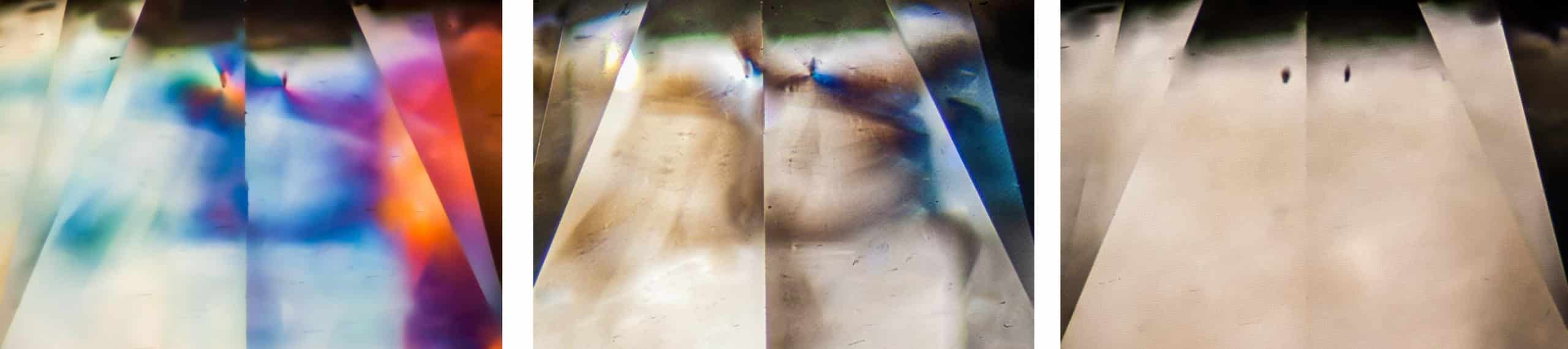
Natural Type Ia(AB) Diamond: Near-colorless round brilliant cut. Not enhanced. This uneven, smudgy appearance is a strong indication of being a “cape series.” Notice the appearance around the small inclusion caused by slight variances in RI (elastic deformation). The field of view is ~3.00mm.
Images courtesy of Randall Lightfoot, CGA.
Images courtesy of Randall Lightfoot, CGA.
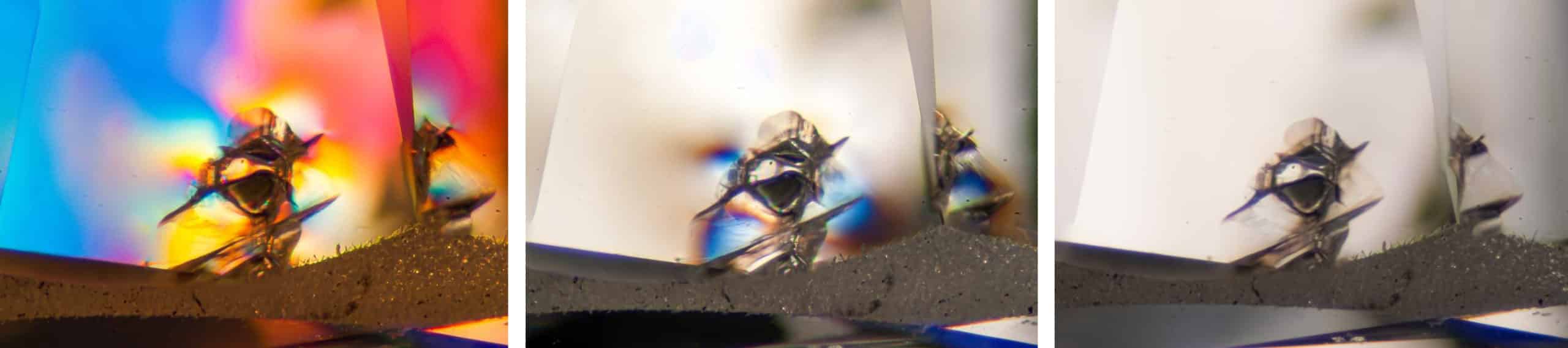
Natural Type Ia(AB) Diamond: Near-colorless round brilliant cut. Not enhanced. Illustrated here is a solid mineral inclusion exhibiting elastic deformation of the surrounding diamond’s crystal lattice. The field of view is ~2.00mm.
Images courtesy of Randall Lightfoot, CGA
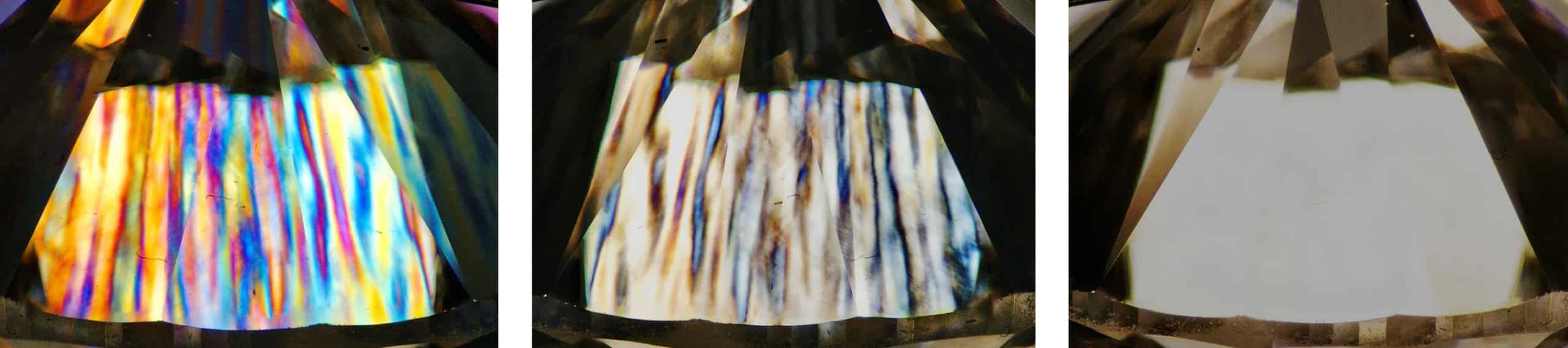
CVD-grown Type IIa Diamond: Near-colorless round brilliant cut. Not enhanced. This is the typical parallel, columnar strain pattern seen in CVD-grown diamonds. The field of view is ~3.00mm.
CVD-grown Type IIa Diamond: Near-colorless round brilliant cut. HPHT-enhanced. Typical cross-hatch/tatami pattern caused by the HPHT treatment. This pattern can also be observed in natural diamonds. This deviates from the typical parallel, columnar pattern we see in CVD-grown diamonds. The field of view is ~3.00mm.
Images courtesy of Randall Lightfoot, CGA.
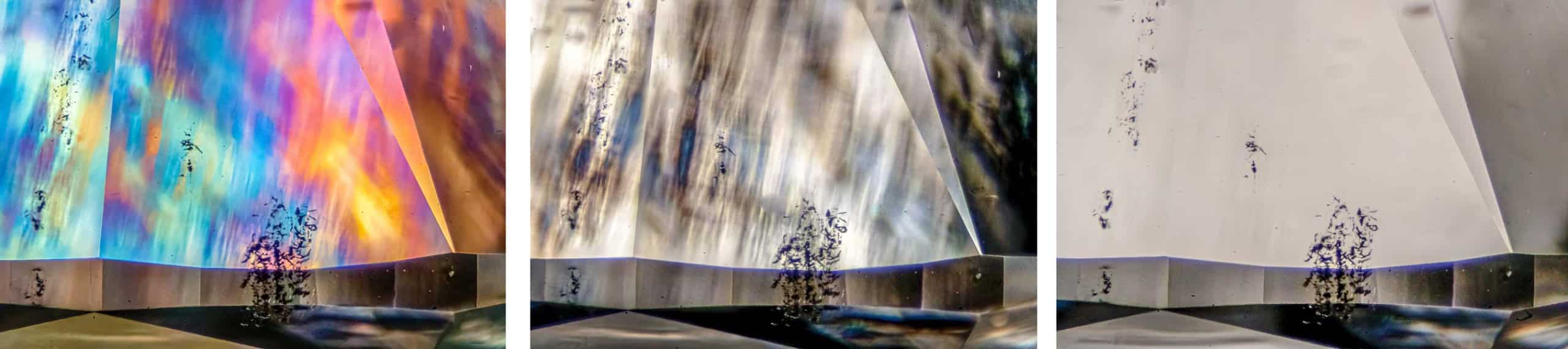
Image property of AGS Laboratories.
All three images here are captured using CPF to demonstrate the differences between natural, CVD-grown, and HPHT-grown diamonds.
Image 1: Natural Type Ia diamond: Typical smudgy appearance – Field of view is ~6.75mm.
Image 2: HPHT-grown Type IIb Diamond: Absence of visible strain – Field of view is ~7.70mm.
Image 3: CVD-grown Type IIa Diamond: Typical distinct columnar growth patterns – Field of view is ~8.45mm.
While CPF is a reliable microscopic method that provides clues to distinguish between most natural and laboratory-grown diamonds, FTIR spectroscopy is required for conclusive identification of diamond type (i.e., Type I or Type II, and their sub-types). Laboratory-grown diamonds are limited to certain diamond types, all of which can be found in natural diamonds as well. Therefore, for a positive determination, advanced testing in a laboratory setting, usually by multiple instruments, is important.
Wade Abel, CG, is the Director of Gemological Services for the AGS Laboratories. He holds his Certified Gemologist® (CG) title from the American Gem Society, Graduate Gemologist Degree from GIA, and a Bachelor’s of Science degree in Business Management from Western Governors University. His gemological career began with AGS Laboratories in May of 2014. Since then, he has been integral in developing new services for AGS Laboratories. He currently oversees all aspects of gemological services at AGS Laboratories and works closely with the AGS membership as committee liaison.
Randall Lightfoot, CGA, crystallized his passion for gemology from years of studying mineralogy and volcanic behavior as an undergraduate. His experience as a diamond grader with the GIA led to his current position as a gemologist appraiser for Christopher’s Fine Jewelry in West Des Moines, IA. He holds a Certified Gemologist® Appraiser (CGA) title from the American Gem Society, is a Registered Gemologist Appraiser with the International School of Gemology, and a Senior Gemologist member with the Accredited Gemologists Association. His specialty is photomicrography and visual identification of gem inclusions.